.png?w=3072&fmt=webp)
Unleashing the
Infinite Possibilities of Live-Cell Imaging,
Pioneering a New Future for Live-Cell Imaging
Unleashing the
Infinite Possibilities of Live-Cell Imaging,
Pioneering a New Future for Live-Cell Imaging

Scroll Down
ABOUT US
关于我们
We innovate for life sciences.
Zircon Optoelectronics (Suzhou) Co., Ltd. is the only high-tech enterprise incubated by the Intelligent Computational Imaging Research Institute of Nanjing University of Science and Technology, dedicated to the industrialization of computational optical microscopy instruments. The team has long been committed to the research and development of computational optical imaging and sensing technologies. Zircon Optoelectronics has developed a range of intelligent computational imaging microscopy products, including label-free intensity diffraction tomography microscopes, reflective and transmissive non-interferometric and multimodal quantitative phase microscopes, and lensless holographic microscopes. From fundamental principles and core technologies to application products, the company possesses fully independent intellectual property rights. Its products have been widely applied in fields such as clinical diagnostics, precision manufacturing, and biomedicine, significantly driving the upgrading of optical microscopy products.
Zircon Optoelectronics (Suzhou) Co., Ltd. is the only high-tech enterprise incubated by the Intelligent Computational Imaging Research Institute of Nanjing University of Science and Technology, dedicated to the industrialization of computational optical microscopy instruments. The team has long been committed to the research and development of computational optical imaging and sensing technologies. Zircon Optoelectronics has developed a range of intelligent computational imaging microscopy products, including label-free intensity diffraction tomography microscopes, reflective and transmissive non-interferometric and multimodal quantitative phase microscopes, and lensless holographic microscopes. From fundamental principles and core technologies to application products, the company possesses fully independent intellectual property rights. Its products have been widely applied in fields such as clinical diagnostics, precision manufacturing, and biomedicine, significantly driving the upgrading of optical microscopy products.
Learn more

核心技术
4th-generation label-free microscopic imaging technology
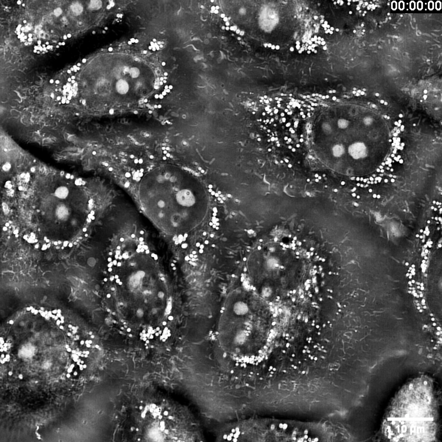
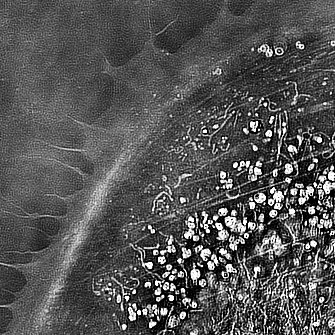
Intensity Diffraction Tomography (IDT) uses the cell’s own refractive index as an “endogenous dye.” By establishing a quantitative link between a through-focal intensity stack and the object’s 3-D refractive-index map, and by engineering an annular, matched-illumination pattern that optimizes the 3-D phase transfer function, IDT reconstructs the volumetric refractive-index distribution from the recorded intensity images through a 4-D optical-transfer-function deconvolution algorithm. The technique not only pushes 3-D diffraction-tomographic resolution to the incoherent diffraction limit, but also delivers high contrast, scattering-robust, and high-axial-sectioning imaging of complex specimens, providing a label-free, quantitative method for live-cell sub-cellular structure imaging and analysis.
Intensity Diffraction Tomography (IDT) uses the cell’s own refractive index as an “endogenous dye.” By establishing a quantitative link between a through-focal intensity stack and the object’s 3-D refractive-index map, and by engineering an annular, matched-illumination pattern that optimizes the 3-D phase transfer function, IDT reconstructs the volumetric refractive-index distribution from the recorded intensity images through a 4-D optical-transfer-function deconvolution algorithm. The technique not only pushes 3-D diffraction-tomographic resolution to the incoherent diffraction limit, but also delivers high contrast, scattering-robust, and high-axial-sectioning imaging of complex specimens, providing a label-free, quantitative method for live-cell sub-cellular structure imaging and analysis.
Learn more

以业务为核心的设计
以业务为核心的设计
Quantitative phase measurement
The light wave reflected or transmitted by the object (object wave) is coherently superposed with a reference wave on the holographic recording plane, forming a hologram that encodes both the amplitude and the phase information of the object wave.
The light wave reflected or transmitted by the object (object wave) is coherently superposed with a reference wave on the holographic recording plane, forming a hologram that encodes both the amplitude and the phase information of the object wave.
联系我们

联系我们

深入研究密切沟通
深入研究密切沟通
Capture holograms from multiple angles
Using annular, matched illumination to optimize the 3-D phase-transfer function, the three-dimensional refractive-index map of the cell is retrieved from the recorded intensity images by deconvolving with the associated four-dimensional optical-transfer-function.
Using annular, matched illumination to optimize the 3-D phase-transfer function, the three-dimensional refractive-index map of the cell is retrieved from the recorded intensity images by deconvolving with the associated four-dimensional optical-transfer-function.
联系我们

超出客户预期
超出客户预期
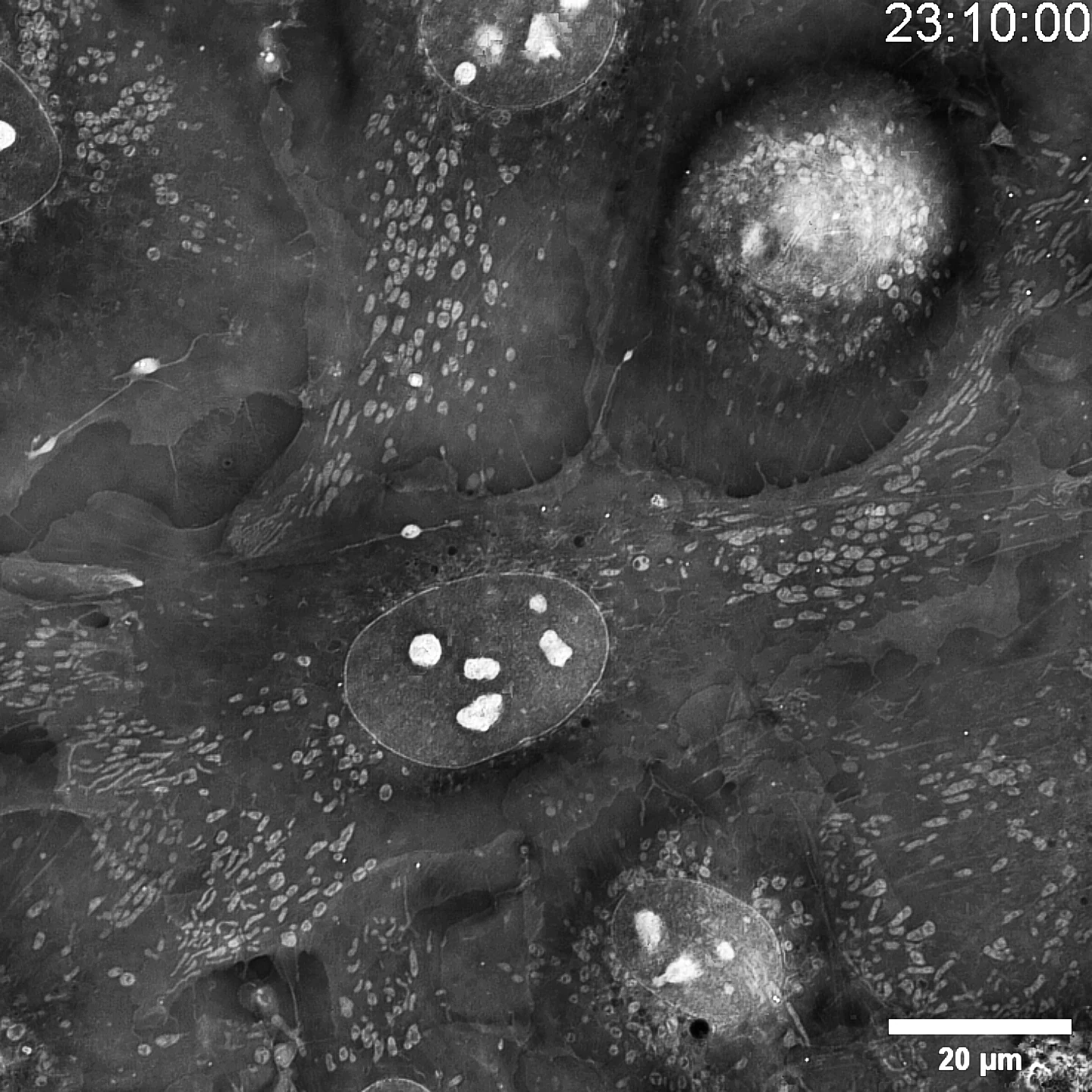
Tomographic 3-D reconstruction
A single shot rapidly delivers 100 focal slices through the specimen, spaced 160 nm apart, letting you watch living cells, sub-cellular features and organelles evolve over time in either 2-D or 3-D—gently, reliably, and with unmatched resolution and speed.
我们所做的一切都是为了实现您战略目标,从本质上讲,我们的设计使您的业务成功的有效方式
联系我们
定量相位测量
定量相位测量
定量相位测量
物体反射或透射的光波(物光)与参考光波在全息记录面上相干叠加,形成全息图。全息图记录了物光的振幅和相位信息。
物体反射或透射的光波(物光)与参考光波在全息记录面上相干叠加,形成全息图。全息图记录了物光的振幅和相位信息。
物体反射或透射的光波(物光)与参考光波在全息记录面上相干叠加,形成全息图。全息图记录了物光的振幅和相位信息。
从多个角度捕捉全息图
从多个角度捕捉全息图
从多个角度捕捉全息图
采用环形匹配照明优化三维相位传递函数,通过相关四维光学传递函数反卷积算法从所记录的强度图像中重建细胞的三维折射率分布。
采用环形匹配照明优化三维相位传递函数,通过相关四维光学传递函数反卷积算法从所记录的强度图像中重建细胞的三维折射率分布。
采用环形匹配照明优化三维相位传递函数,通过相关四维光学传递函数反卷积算法从所记录的强度图像中重建细胞的三维折射率分布。
层析重构三维
层析重构三维
层析重构三维
单次成像即可快速获得目标样品100层不同焦面图像信息,三维衍射层析间距达到160nm,您可2D和3D形式观察活体样品中的细胞、亚细胞以及细胞器结构随时间变化的情况,并以更温和可靠,且更出色的分辨率和成像速度完成观察。
单次成像即可快速获得目标样品100层不同焦面图像信息,三维衍射层析间距达到160nm,您可2D和3D形式观察活体样品中的细胞、亚细胞以及细胞器结构随时间变化的情况,并以更温和可靠,且更出色的分辨率和成像速度完成观察。
单次成像即可快速获得目标样品100层不同焦面图像信息,三维衍射层析间距达到160nm,您可2D和3D形式观察活体样品中的细胞、亚细胞以及细胞器结构随时间变化的情况,并以更温和可靠,且更出色的分辨率和成像速度完成观察。
03
产品
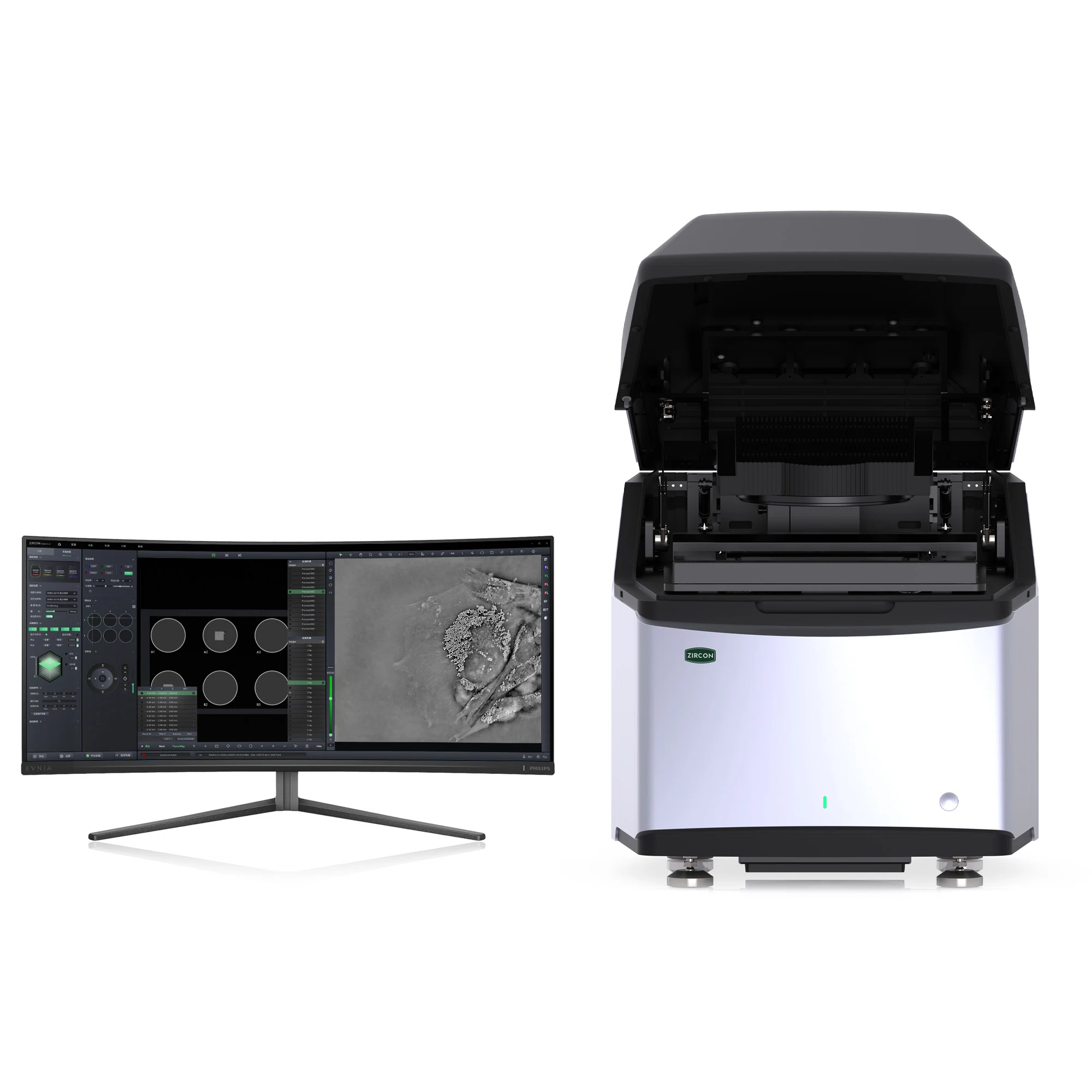
SC3000
无标记活细胞显微成像系统
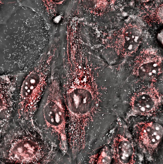
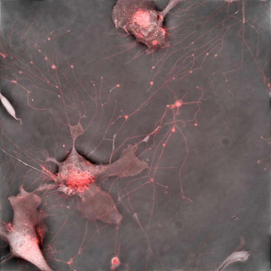
Zircon Optoelectronics SC3000 harnesses Intensity-Diffraction-Tomography (IDT) to visualize sub-cellular architecture in living cells for unlimited time-lapses—no stains, no labels. IDT pushes 3-D tomographic resolution to the incoherent diffraction limit and, even in thick, scattering specimens, delivers high-contrast, scatter-immune, high-axial-sectioning imaging, giving researchers a truly quantitative, label-free assay of sub-cellular dynamics. Integrating 4th-generation non-interferometric label-free imaging, high-resolution fluorescence, and a full live-cell workstation, SC3000 supports studies of cell–cell interactions, migratory behavior, organelle organization and thick-tissue imaging, providing an untouched, high-fidelity window into living biology.
Zircon Optoelectronics SC3000 harnesses Intensity-Diffraction-Tomography (IDT) to visualize sub-cellular architecture in living cells for unlimited time-lapses—no stains, no labels. IDT pushes 3-D tomographic resolution to the incoherent diffraction limit and, even in thick, scattering specimens, delivers high-contrast, scatter-immune, high-axial-sectioning imaging, giving researchers a truly quantitative, label-free assay of sub-cellular dynamics. Integrating 4th-generation non-interferometric label-free imaging, high-resolution fluorescence, and a full live-cell workstation, SC3000 supports studies of cell–cell interactions, migratory behavior, organelle organization and thick-tissue imaging, providing an untouched, high-fidelity window into living biology.
Learn more
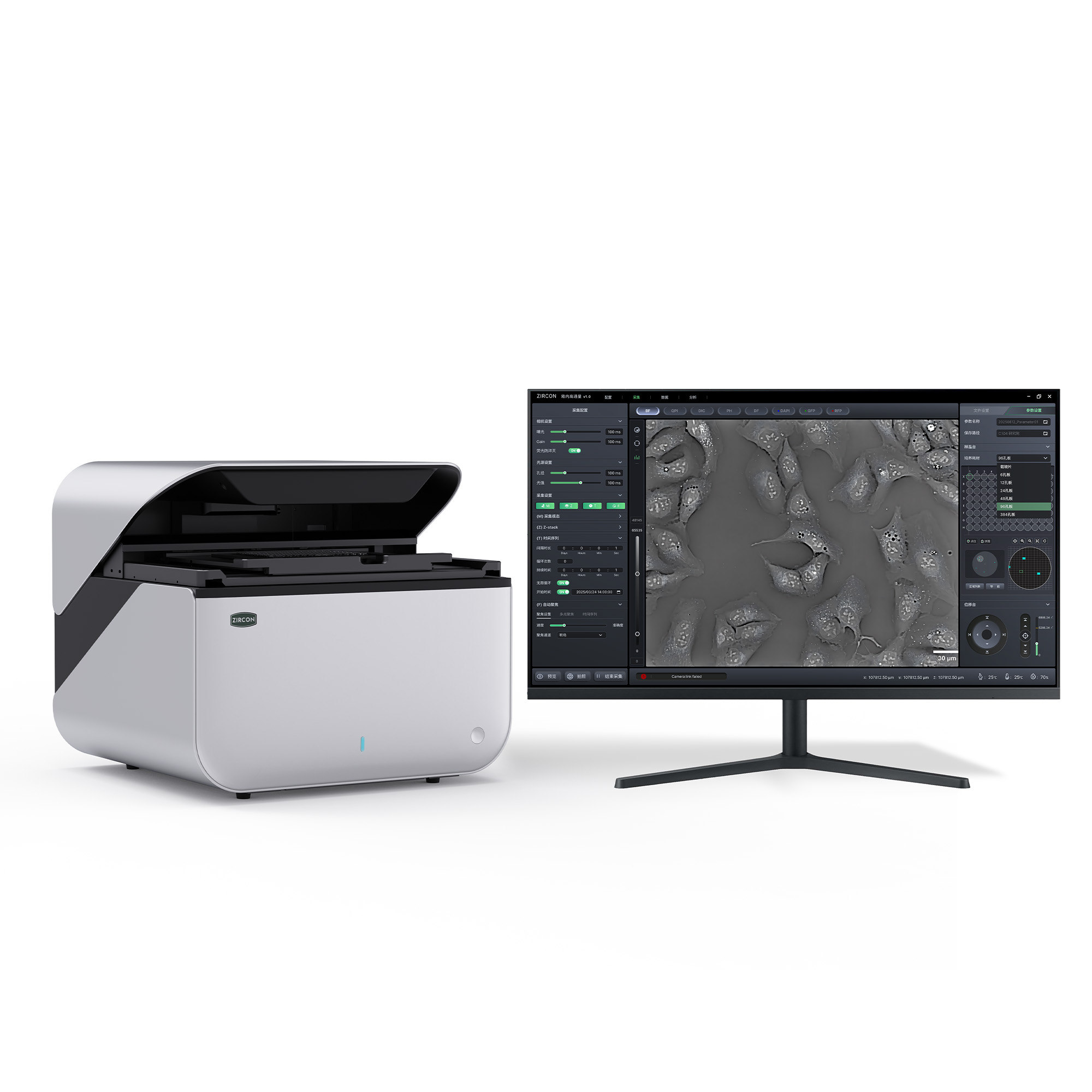
全自动多模态显微成像系统
C600
Zircon Optoelectronics C600 leverages proprietary, non-interferometric, label-free refractive-index imaging to deliver 260 nm sub-cellular resolution with full 96-well-plate high-throughput scanning—ideal for long-term single-cell tracking and organ/embryo-development monitoring. The platform revolutionizes workflow by integrating six core modalities—bright-field, dark-field, phase-contrast, DIC, label-free and fluorescence—into a single, button-switchable optical path with no mechanical flipping, boosting experimental throughput. Built-in 2-D/3-D fluorescence deconvolution fused with large-model image segmentation enables high-precision single-cell quantification. A motorized stage and multi-dimensional analytics suite deliver an all-in-one solution for high-content screening and cytotoxicity studies.
Zircon Optoelectronics C600 leverages proprietary, non-interferometric, label-free refractive-index imaging to deliver 260 nm sub-cellular resolution with full 96-well-plate high-throughput scanning—ideal for long-term single-cell tracking and organ/embryo-development monitoring. The platform revolutionizes workflow by integrating six core modalities—bright-field, dark-field, phase-contrast, DIC, label-free and fluorescence—into a single, button-switchable optical path with no mechanical flipping, boosting experimental throughput. Built-in 2-D/3-D fluorescence deconvolution fused with large-model image segmentation enables high-precision single-cell quantification. A motorized stage and multi-dimensional analytics suite deliver an all-in-one solution for high-content screening and cytotoxicity studies.
Learn more
案例
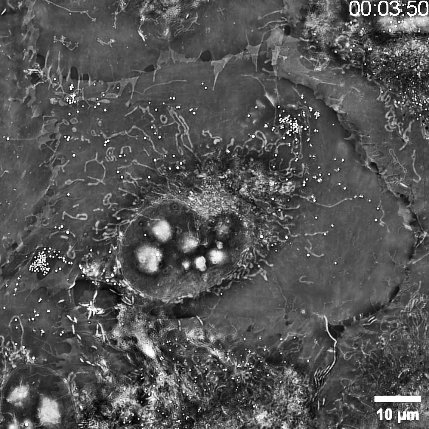
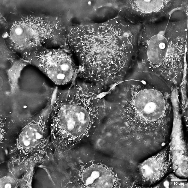
Imaging Gallery
Imaging Gallery
新闻
NEWS
更多新闻
合作咨询
致力于为国内外品牌和企业提供极致的数字创意服务,为重塑新一代互联网用户体验,我们的奇思妙想帮助企业率先触摸未来。我们的工作永远以客户为中心,从策略、创意、设计,到可扩展的品牌数字化、商务、移动和数字平台,我们愿始终于合作客户并肩作战,联合创新,共同实现价值。

查看更多
Learn more

查看更多
Learn more

查看更多
Learn more
06
合作单位
Collaborating Universities & Research Institutions


导航1
导航2
导航3